Desert ImagingM R I / M R A
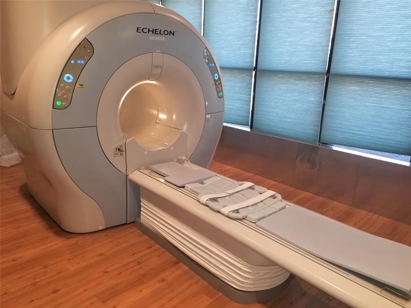
M R I
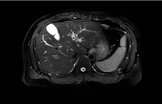
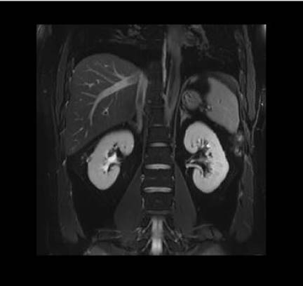
An MRI scan, also called magnetic resonance imaging uses a strong magnet and radio waves to align water molecules in the area being imaged. Since our bodies are made up of mostly water and each organ has different quantities of water in it, the images acquired show differences in normal and abnormal anatomy.
Digital geometry
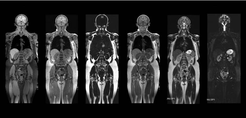
Digital geometry processing is used to generate a 3D (3 dimensional) image of the inside of the object from a large series of 2D (two-dimensional) images. These 3D images are put together to generate the image of a particular organ.
( Westside: 122 West Castellano Drive with 3-Tesla and 1.5-Tesla systems - Eastside: 1727 Lee Trevino with a 1.5-Tesla system )
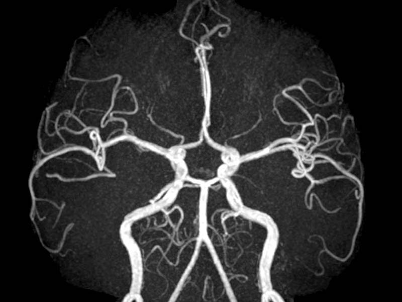
M R A
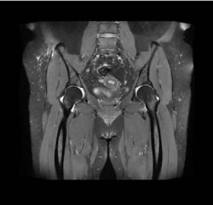
An MRA is a magnetic resonance angiography which generates pictures of the arteries to evaluate them for abnormal narrowing or aneurysms. MRA is often used to evaluate the arteries of the neck and brain, the thoracic and abdominal aorta, the renal arteries, and the legs called a run-off.
Both MRI and MRA images can generate a 3D image which can help the radiologist find abnormalities.

Discover what ultra wide bore 3T MRI can do for you.
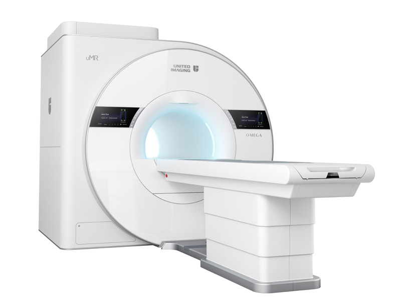
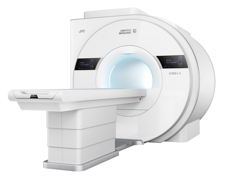
3-Tesla MRI
More comfortable. Higher quality images. MRls in half the time. Desert Imaging now offers state-of-the-art 3-Tesla MRI exams on our new ultra-wide bore uMR OMEGA™ system that delivers unrivaled speed, quality, and comfort to El Pasoans.
Our new MRI will increase diagnostic confidence, reduce patient stress/claustrophobia, and expand access to more patients in our community.
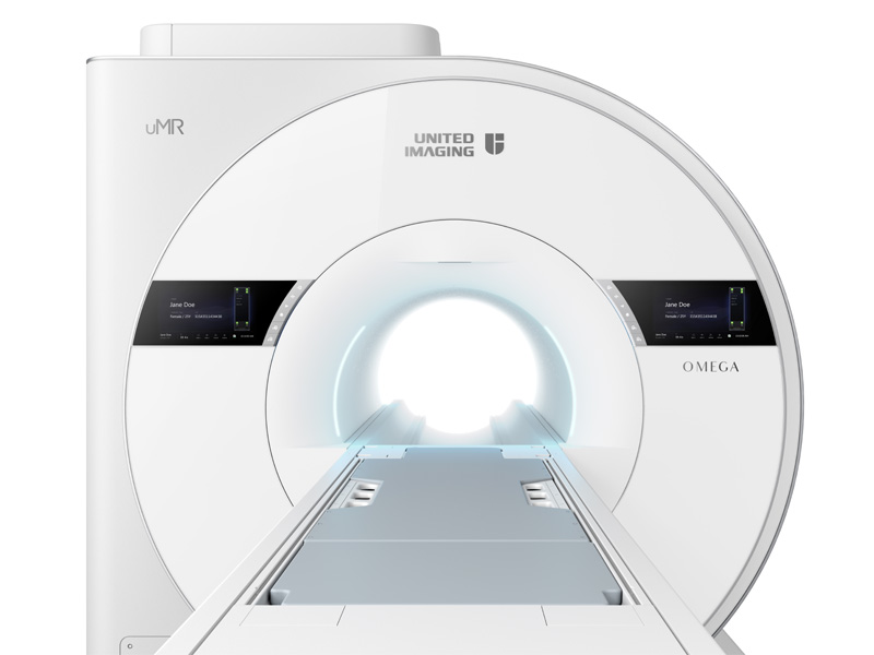
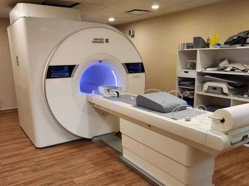
World Class 3T MRI
COMFORT
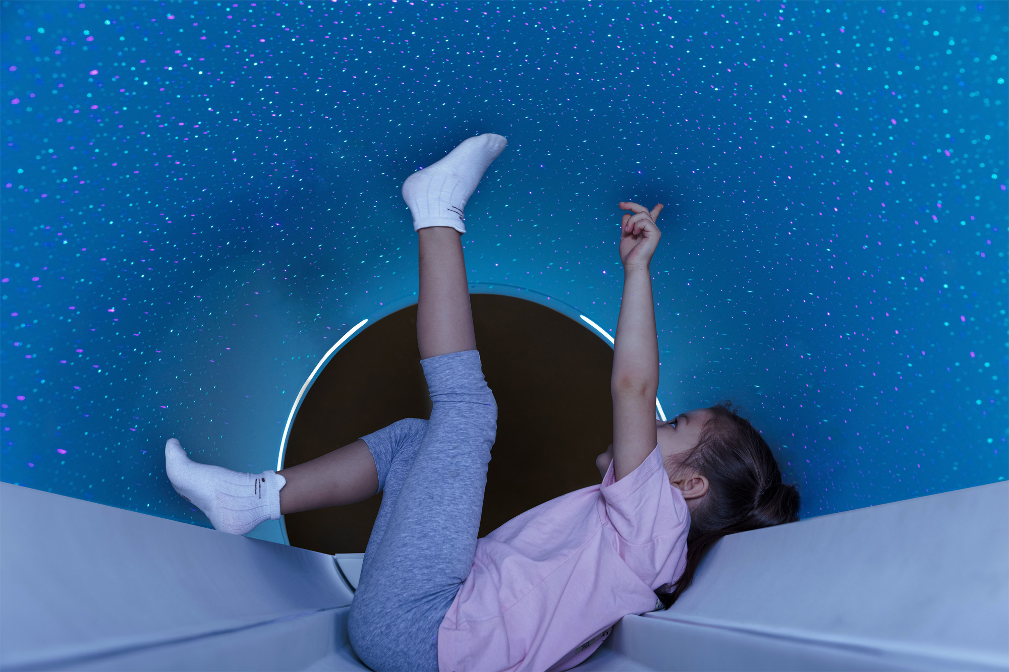
- Widest bore on market (75 cm) with a starlight ceiling
- Headset provides music and communication
- Free breathing capability with ultra short breath holds
2X FASTER EXAM TIMES
- 50% shorter examinations
- Less Claustrophobia, Stress & Anxiety
- AI-Empowered Compressed Sensing (uAI ACS)
EXCEPTIONAL IMAGE QUALITY
- Best-in-class homogeneity and gradient performance
- World’s largest field of view (60x60x50 cm)
- Motion insensitive sequences for clearer images
To schedule an appointment with Desert Imaging, simply call:
(915) 577-0100
Frequently asked questions
Did you know?
MRI / MRA
Both MRI and MRA images can generate a 3D image which can help the radiologist find abnormalities.
- Pacemaker
- Pregnancy
- Claustrophobia
- History of kidney problems
- Skin tattoos
- Neurostimulators (TENS-unit)
- Implanted drug infusion device (i.e., insulin pump)
- Exposure of metal fragments to your eye
- Artificial heart valves
- Aneurysm clips
- Cochlear implants
- Metallic implants and prosthesis
- Vascular stent or stent graft
- History as a metal worker
- Shrapnel or bullet wounds
- Dorsal column stimulators
- Allergy to iodine, or gadolinium
- History of diabetes
- Other conditions you believe to be relevant
![]()
- Arrive at least 15 minutes before your scheduled appointment time.
- Bring all health insurance cards and information.
- Notify us if any of your information has changed
- Wear loose, comfortable clothing
- Leave valuable items at home